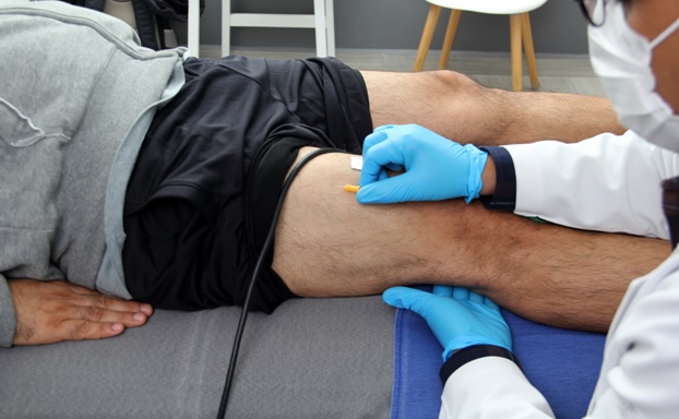
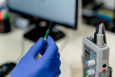
Electromyography, or EMG, traditionally significant for its use in diagnosing a wide range of neuromuscular disorders, is only growing in importance for its use in researching control of robotic and prosthetic mechanisms.1
As useful as electromyography promises to be in ongoing medical and engineering applications, EMG signals can be compromised by a wide range of impediments, known as artifacts, which can severely complicate the interpretation of the recording.
Even with the use of high-quality EMG electrodes and equipment, and when following accepted best medical practices, artifacts in EMG recordings can still present a significant obstacle to accurate readings. The purpose of this short article is to expose some of the causes of these artifacts, as well as how to minimize them.
The Causes of Artifacts and Signal Interference in Electromyography
One of the most obvious causes of artifacts in electromyography is the presence of inherent competing signals from the electrical equipment used. Unfortunately, this cannot be eliminated. It can be detected, and, at best, mitigated by using high-quality EMG electrodes and equipment.2 Ensuring the skin is clean, and the needles inserted (or sEMG electrodes attached) properly, can help with this. Do not allow EMG lead wires to contact metal surfaces or become coiled. If you can identify a strong source of electromagnetic interference (EMI), if possible, move it. A small relocation can have a big impact on the magnitude of the interference.
Motion artifacts are another adverse impactor of EMG signal quality. However, despite the name, they are not caused by the motion of the patient (which is inherent in some of these tests, anyway) but by irregularities in the electrode interface or cable.
Another common cause of artifacts in EMG signal recording is a result of the inherent instability of the baseline being a proper ground very important for the procedure. Preparing the Patient
Depending on the nature of the EMG test, you may want to inform your patient very carefully how to prepare for the test.
Instruct your patients not to ingest caffeine for a period of at least 6 hours before the test. Non-caffeinated teas and coffees are acceptable, but foods and drinks containing them (like soda) should be avoided.

Identifying and Ameliorating the Causes of EMG Artifacts
There are a variety of methods applied to decontaminating the signal of an EMG recording without compromising its fidelity.
One common method for removing the influence of noise from an EMG recording is the use of conventional digital filters, of which there are three common types: low-pass filters, high-pass filters, and band-stop filters.
Low-pass filters remove any signal with a frequency of 500 Hz, which would be considered noise in an sEMG, although the cutoff frequency can be adjusted.
The use of high-pass filters to remove artifacts from surface electromygraphy (sEMG) signals is more complicated as many of the contaminants may have overlapping frequencies. Therefore, the cutoff frequency must be set according to the application and the muscle studied.
Band-stop/notch filters are used predominantly to combat the effects of PLI, or power-line interference.
Gating and clipping are also used to eliminate competing signals from the EMG recording based on time range. Gating is the simplest method, in which a specific time series (containing an artifact) is isolated and removed from the recording. Unfortunately, gating does not allow for a continuous EMG signal and presents the risk that valuable electrical data may be overlooked.1
As for clipping, it basically eliminates a set of amplitudes based on frequency; the only problem is clipping can be more deleterious to the overall signal than the interference itself.1
Another method for removing interference is called the “adaptive estimation of the interference signal by filtering the raw signal”.1 Effectively, this method subtracts the interference signal from the raw signal to estimate the EMG signal. This does, of course, require an accurate determination and estimation of the interference signal itself.
Adaptive noise cancellers can also be used to varying effect to eliminate electromagnetic interference. Also known as ANCs, these help eliminate some of the problems associated with identifying the competing frequency. Identifying and removing the wrong frequency can end up exacerbating the artifact instead of diminishing it.
Adaptive noise cancellers use a noise reference signal, captured at the same time as the EMG signal but through a different channel. This reference signal is then modified using an adaptive algorithm that estimates the noise, which is then subtracted from the raw signal, which should, in turn, yield a higher-quality EMG signal.

High-Quality EMG Electrodes
This is only a very small cross-sectional sampling of the different techniques used to assess EMG signals in an attempt to identify and remove artifacts. Part of capturing a high-quality EMG recording begins with the use of quality EMG electrodes and other electrical equipment.
LifeSync Neuro is your source for high-quality EMG electrodes, including but not limited to color-coded, single-patient-use, disposable electrodes, as well as concentric needle electrodes and monopolar electrodes. We carry interface cablesin both shielded and unshielded configurations.
We also carry injectable needle electrodes with attached leads, cables, and offer bulk discounts. Contact us for more information if you have any questions about any of our products or policies.
Sources
- Boyer M, Bouyer L, Roy JS, Campeau-Lecours A. Reducing Noise, Artifacts and Interference in Single-Channel EMG Signals: A Review. Sensors (Basel). 2023 Mar 8;23(6):2927. doi: 10.3390/s23062927. PMID: 36991639; PMCID: PMC10059683.
- Raez MB, Hussain MS, Mohd-Yasin F. Techniques of EMG signal analysis: detection, processing, classification and applications. Biol Proced Online. 2006;8:11-35. doi: 10.1251/bpo115. Epub 2006 Mar 23. Erratum in: Biol Proced Online. 2006;8:163. PMID: 16799694; PMCID: PMC1455479.

